7.2 Animal Tissue:
The tissues in the body of animals are called Animal Tissue.Savinay Kumar JC
You may be surprised to know that all complex animals consist of only four basic types of tissues.
These tissues are organized in a specific proportion and pattern to form an organ like stomach, lungs, heart and kidney.
In animals, tissues are grouped into four types based on their functions as follows;
Epithelial Tissue
Connective Tissue
Muscular Tissue and
Neural Tissue
7.2.1 Epithelial Tissue: (Gr; Epi = on; thelial = nipple)
An epithelium is a tissue composed of one or more layers of cells that cover the body surface and line its various cavities.
General Function: It serves for protection, secretion and excretion.
Types of Epithelial tissues:
Based on number of cell layers, epithelial tissues divided into two types as follows;
Simple Epithelial tissue and
Compound Epithelial tissue.
7.2.1.1 Simple Epithelial Tissue:
It is composed of a single layer of cells.
Major Function: It is a lining for body cavities, ducts and tubes.
Other functions like diffusion, secretion, absorption and movement of the particles.
Types of Simple Epithelial Tissue:
Based on shape of epithelial cell, it is further divided into four types as follows;
Squamous Epithelium
Cuboidal Epithelium
Columnar Epithelium and
Ciliated Epithelium.
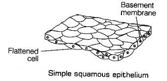
7.2.1.1.1 Squamous Epithelium:
It is made up of flattened cells with irregular boundaries. Its nuclei are located at the center.
Ex: Walls of Blood Vessels, air sacs of lungs.
Function: forming a diffusion boundary.
7.2.1.1.2 Cuboidal Epithelium:
It is composed of cube-like cells. Its nuclei are located at the center.
Ex: ducts of glands and tubular parts of nephrons.Savinay Kumar JC
Function: secretion and absorption.
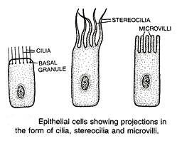
The epithelium of proximal convoluted tubules (PCT) of nephrons in the kidney has microvilli.
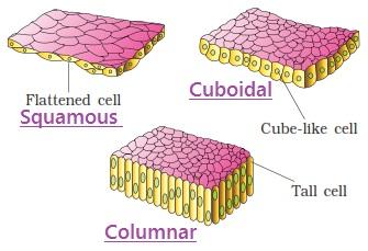
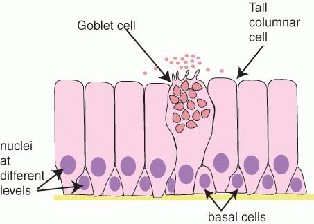
7.2.1.1.3 Columnar Epithelium:
It is composed of tall and slender cells (column like cells).
The nuclei of the columnar cells are located at the base and in few cases center.
Free surfaces may have microvilli.
Ex: lining of stomach and intestine.
Function: secretion and absorption.
7.2.1.1.3 Ciliated Epithelium:
It is a modified Cuboidal or columnar epithelium cell that bears cilia on their free surfaces.
Ex: inner surface of hollow organs like bronchioles and fallopian tubes.
Function: to move particles or mucus in a specific direction over the epithelium.
7.2.1.2 Compound Epithelial Tissue:
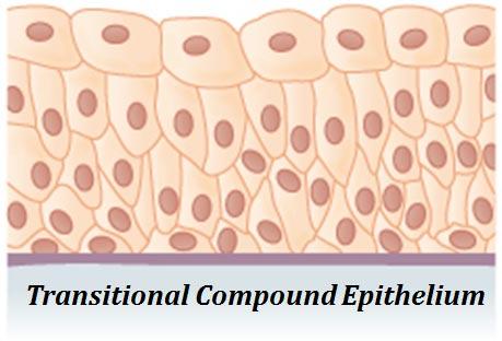
It is composed of more than one layer of cells.
Ex: surface of skin, buccal cavity, duct of salivary gland and pancreatic duct.
Major Function: protection against chemical and mechanical stress.
Other functions like secretion and absorption are observed in a few cases.
7.2.1.3 Glandular Epithelium:
Some of the columnar or cuboidal cells specialized for secretion is called glandular epithelium.Savinay Kumar JC
Types of glandular epithelium: Based on number of epithelial cells participate in secretion, glandular epithelium divided into two types as follows;
Unicellular Glandular Epithelium and
Multicellular Glandular Epithelium
7.2.1.3.1 Unicellular Glandular Epithelium:
It is also called simple gland or goblet cells.
It consists of isolated glandular cells (one cell) in the epithelium.
Ex: Alimentary canal.
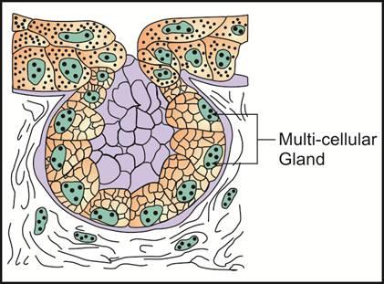
7.2.1.3.2 Multicellular Glandular Epithelium:
It consists of many cells for secretion.
Ex: Salivary Gland.
Note: According to the Greek language Exo = outside; Endo = inside; and crine = secrets.
7.2.1.3.3 Difference between Exocrine and Endocrine Glands:
7.2.2 Connective Tissue:
Connective Tissue is also known as binding tissue because of their special function of linking and supporting other tissues or organs of the body.
The connective tissues are most abundant and widely distributed in the body of complex animals.
The cells of the connective tissue secrete fibers of structural proteins called collagen or elastin.
But blood is an exception; in which no structural fiber is secreted.
The structural fibers provide strength, elasticity and flexibility to the tissue.
The cells of the connective tissue also secrete modified polysaccharides. These polysaccharides accumulate between cells and fibers and act as a matrix (ground substances).
Ex: Cartilage, Bone, Blood, Areolar, Adipose and Tendons.Savinay Kumar JC
Types of Connective Tissue:
Based on their arrangement and function, connective tissue is divided into three types as follows;
Loose Connective Tissue
Dense Connective Tissue and
Specialized Connective Tissue
7.2.2.1 Loose Connective Tissue:
Loose connective tissue has cells and fibers loosely arranged in a semi-fluid substance.
Examples for Loose Connective Tissue are as follows;Savinay Kumar JC
Areolar Tissue and
Adipose Tissue.
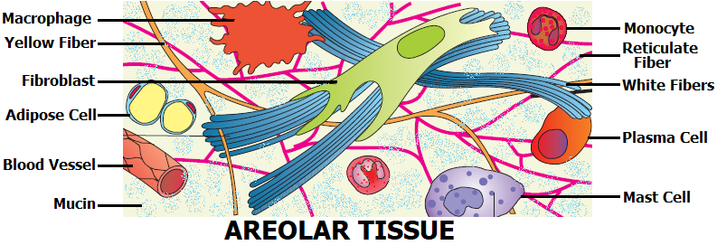
Ex 1: Areolar Tissue:
Areolar Tissue is a type of loose connective tissue, it is present beneath the skin and it supports the framework for epithelium.
It consists of Mucin (matrix/fluid), three types of fibers and five types of cells.
Note:Savinay Kumar JC
White Fibers = Collagen 1 Fibers
Yellow Fibers = Elastin Fibers
Reticular Fibers = Collagen 3 fibers
Extra Information:
White Fibers: It contains a greater proportion of white inelastic fibers. Function = mechanical strength.
Yellow Fibers: It is made up of elastic fibers. Function = Stretch without damaging tissue.
Reticular Fibers: It is made up of type 3 collagen.Savinay Kumar JC
Function = it acts as a supporting mesh in soft tissue.
Fibroblasts: They are large, flat, highly branched cells.
Function = secrete and maintain the fibers.
Plasma Cells: They are oval in shape, agranular cytoplasm and small nucleus. Function = produce clotting factor and antibodies.
Adipose: They are the fat filled cells. Function = provide energy to the body.
Macrophages: They are amoeboid in shape. Function = they defend and ingest (eating) the infectious microorganism.
Mast Cells: They are the large cells that have a spherical nucleus with granular cytoplasm.
Function = secretion of serotonin, heparin and histamine.
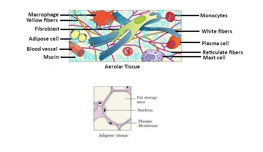
Ex 2: Adipose Tissue:
Adipose tissue is another type of loose connective tissue located mainly beneath the skin.
Functions:Savinay Kumar JC
The cells of this tissue are specialized to store fats.
The excess of nutrients which are not used immediately are converted into fats and are stored in this tissue.
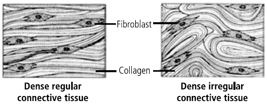
7.2.2.2 Dense Connective Tissue:
It is made up of Fibers and Fibroblasts in their matrix.
Types of Dense Connective Tissue: Based on arrangement of white fibers and fibroblast cells, it is divided into two types as follows;
Dense Regular Connective Tissue and
Dense Irregular Connective Tissue
7.2.2.2.1 Dense Regular Connective Tissue:
Orientation of fibers and fibroblast shows a regular pattern called Dense Regular Connective Tissue.
Ex: Tendons (attach skeletal muscles to bones) and Ligaments (attach bones to other bones)
7.2.2.2.2 Dense Irregular Connective Tissue:
Orientation of fibers and fibroblast shows an irregular pattern called Dense Irregular Connective Tissue.
Ex: Present in the skin.
7.2.2.3 Specialized Connective Tissue:
The connective tissue, which performs other than binding or linking or connecting. This kind of connective tissue is called specialized connective tissue.
Ex: Bone, Cartilage and Blood.
Difference between Cartilage and Bone:
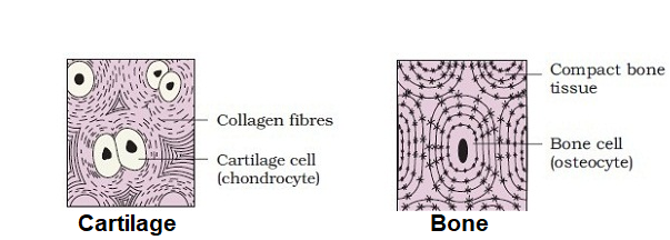
Note:
The Cartilage and bone cells are present in the spaces called lacunae. Cartilage and bone are both structural materials.
Blood:Savinay Kumar JC
It is a fluid connective tissue.
It contains plasma, blood cells and platelets.
Blood is the main circulating fluid which helps in transport of various substances.
7.2.3 Muscle Tissue:
This is a tissue that helps in the movement of the various body parts.
The muscular tissue is generally attached to the bones and thus helps in movement.
Muscle cells are called the myocytes.
They have myofibrils which are composed of actin and myosin / myofilaments.
They slide past each other, which produces tension, thus changing the shape of the myocyte.
In a single muscle tissue, there are many myocytes present. When the muscle fibers contract and relax, movement occurs.
Contraction of the muscle fibers is in response to a stimulus.
Types of Muscle Tissue: Based on the shape and function, Muscle tissue is divided into three types as follows;
Skeletal Muscle
Smooth Muscle and
Cardiac Muscle
Difference between Skeletal, Smooth and Cardiac Muscles:
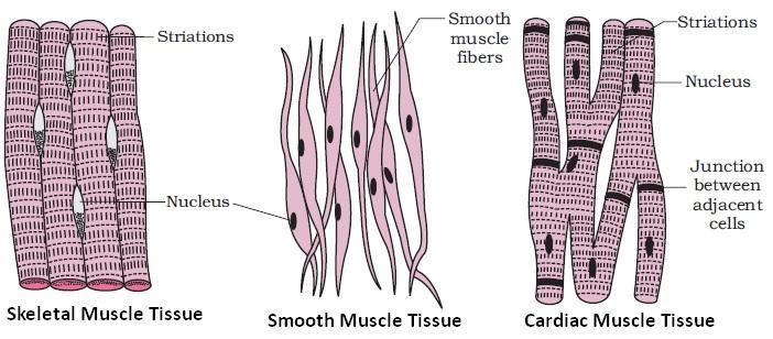
Note: Cardiac Muscle Tissue is also known as communicating Muscles because between the cells contain communication junctions and Intercalated discs.
7.2.4 Neural Tissue:
It is also known as Nervous or Nerve Tissue.
The units of neural tissue are Neurons
It is the main tissue component of the two parts of the nervous system; the central nervous system (CNS), and the peripheral nervous system (PNS).
It regulates and controls bodily functions and activity.
It is made up of cells called neurons and neuroglia.Savinay Kumar JC
Neuroglia: A delicate connective tissue which supports and binds together the nerve tissue in the Central Nervous Tissue.
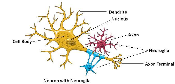
Neuroglia functions: It maintains homeostasis, forms myelin, provides support and protection for neurons.
7.3 Cell Junction:

All animal tissues are held together with little intercellular material between cells known as cell junction.
Cell junctions are intercellular connections between the plasma membranes of adjacent cells in animal tissues.
Cell junction if observed in epithelium is called epithelium junction.
7.3.1 Types of Cell Junction:
Based on their functions, it is divided into three types as follows;Savinay Kumar JC
Tight Junction: It helps to stop substances from leaking across a tissue.
Adhering Junction: It performs cementing to keep neighboring cells together.
Gap Junction: It facilitates the cells to communicate with adjoining cells.


No comments:
Post a Comment